 |
||
| HOME | ||
|
||
| Acute Myelogenous Leukemia (AML) |
||
|
||
| Other Leukemia Types (ALL / CLL / CML / HCL) |
||
|
||
| Myelodysplastic Syndrome | ||
|
||
| Symptoms and Diagnosis | ||
|
||
| Leukemia Treatment Options | ||
|
||
| " Chemotherapy | ||
|
||
| " Blood Stem Cell Transplants | ||
|
||
| " Radiation and Surgery | ||
|
||
| " Chemo Side Effects | ||
|
||
| " Clinical Trials Info | ||
|
||
| " Coping with Leukemia | ||
|
||
| " What to Ask Your Doctor | ||
|
||
| Financial Assistance | ||
|
||
| At Risk Jobs/Exposure | ||
|
||
| Leukemia Resources | ||
|
||
| Survivor's Story | ||
|
||
| Leukemia News | ||
|
||
|
Search for information:
|
||
 |
Blood Cancer &
|
 |
 Leukemia Cancer Information
|
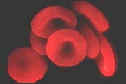
Leukemia Cancer News October 2004 The Marrow of Life By Angilee Shah, AlterNet But a sudden attack of headaches and breathing problems altered the 22-year-old's plans. In a matter of hours, she discovered that she had acute myelogenous leukemia, or AML, a cancer that affects the blood and bone marrow. Today, instead of rushing to the New York subway every morning to get to a newsroom, she stays within the walls of the City of Hope National Medical Center in Southern California to be monitored and medicated. She was admitted to the hospital on Aug. 15 for a second round of chemotherapy, but she has still not gone into remission. A bone marrow transplant is the only treatment option left for Sagarika. So she waits in the hospital, where she will most likely remain until a donor with her tissue type can be found. "Now chemotherapy is not an option," says Sagarika's father, Anand Savur. "Her only hope is a bone marrow transplant," he says, adding, "Time is critical now." AML produces leukemia cells quite rapidly. Without any treatment, the disease can be fatal in a few months. Sagarika's battle is an especially difficult one; not just because of the pain caused by leukemia or of the side effects of chemotherapy but because Sagarika is South Asian. A patient must find a bone marrow donor whose tissue type matches his or her own. The highest chance of a match comes first from within the patient's immediate family, and second from someone within the same ethnic group the common genes allow for similar tissue types. AML is a type of cancer in which the patient cannot produce enough healthy blood cells because of unhealthy bone marrow, the tissue inside the bone that creates different types of blood cells. People with leukemia do not have enough healthy marrow cells, more commonly known as blood stem cells (not to be confused with embryonic stem cells, which come from human embryos). Blood stem cells are blank, not-yet-mature cells that have the capability of becoming platelets, red or white blood cells. In individuals with leukemia, the unhealthy blood stem cells create too many immature white blood cells, which in turn become leukemia cells. These leukemia cells make a patient susceptible to infection, and can cause bone pain, fever, and a host of other symptoms. For Sagarika, this means that she can only seek a match from the less than 1 percent of registered donors in America who are South Asian. Because the United States has the only national, centralized registry that includes a significant number of South Asians (South Asian countries, like India and Pakistan, do not have centralized registries), Sagarika and 35 other South Asian Americans like her are hoping that more South Asians will join the national registry to increase their chances of finding a match. In 2003, the national registry was only able to match about 2,000 minorities, out of over 15,000 matches in total. The National Marrow Donor Program (NMDP) organizes and maintains the registry; of the 5.4 million people in its database, only 64,000 are of South Asian descent. Groups like the South Asian Marrow Association of Recruiters, or SAMAR and Asians for Miracle Marrow Matches or A3M, are stepping in to fill the minority void. Both organizations hold drives and programs to raise awareness around the issue. But it is not easy to convince people to register. "It requires a lot of pre-education," says Enisha Narang, South Asian Outreach and Recruitment Coordinator for A3M. The NMDP says that, on average, a person needs to hear about bone marrow donation seven times before even considering registration. The relative success of drives, therefore, is not just about how many people register, says Narang. "Even if we don't recruit a lot of donors, we've gotten information out." And getting the word out seems to be helping. Narang says A3M targets South Asians where they live. "We organize drives, she says, "at all different sorts of events wherever South Asian people gather," including temples, mosques, gudwaras, and even in private homes. SAMAR uses the same strategy of community and volunteer-based organizing. Dr. Asif Amirali, a SAMAR volunteer in New York City, says that they have been successful, since their inception in 1992, in directly recruiting from start to finish 35,000 South Asians to register and helped to register 20,000 others. The rate of registration continues to rise. SAMAR is also focusing some of its efforts in creating a national registry in India, which would be available for South Asians both on the subcontinent and in America. They face significant hurdles though. "It's not easy to get the [Indian] government to commit resources to something like bone marrow registry," says Amirali. So SAMAR is looking for money from private and public sources to build the special labs and pay for testing. Because of the time it will take to begin the registry, plus the time is will take to build a list of donors, Amirali says, "It's going to take years to have a fully functioning registry [in India]." Mrinmayee Kulkarni, 25, is one of the SAMAR volunteers who make drives and recruitment possible in America. Her work is driven by immediate necessity; Sagarika is one of Kulkarni's best friends. Their families were neighbors in India, so they have known each other since they were small children and have kept in touch ever since. When Kulkarni learned of her friend's illness, she immediately took action. "I had initially contacted SAMAR to get tested," says Kulkarni, but she realized that the cure is much more complicated than a single person deciding to register. "They [patients] don't have any other cure. Sagarika is undergoing chemotherapy but that's just buying time," says Kulkarni. Potential Donors on the National Registry SOURCE: Patrick Thompson, Senior Public Relations and Media Outreach Coordinator, National Marrow Donor Program NMDP explains in its literature the process of bone marrow registration and donation. In the initial testing, a small amount of blood is drawn from the finger of the potential donor. If the donor is found to match a patient, a few more tests are run and then if the donor decides to continue the donor chooses if he or she wants to draw marrow cells from the hipbone or the blood. According to the NMDP web site, the marrow procedure takes about one hour and is done under general or local anesthesia, which is again, the donor's choice. Dr. Amirali says a common misperception is that the marrow cells are taken from the back or spine, when in actuality, the needle goes into the hipbone on the side of the body a much less painful experience. "A six-month-old child has donated through the hipbone so any one of us can do it," says Amirali. To donate from the blood stream, the donor must receive special injections for a few days. In the actual procedure, the blood is drawn through the arm and then run through a machine. In that machine, marrow cells are separated out and stored away in a process called aphaeresis. The blood, without marrow cells, is then returned to the donor's body through the other arm. Donors recover in one or two days and experience manageable aches. "Most people are able to go back to work the next day or the day after," says Amirali, "and any pain is manageable with Tylenol or Advil." Kulkarni says, "The donation is not painful or life-threatening as people believe. It's not like the surgery it used to be 10 years ago." Furthermore, for minorities, registrations can be done free of charge through groups like SAMAR. The patients receiving the transplants cover any subsequent donations. The cost of the procedures, however, is much less than the cost of not being able to find a match. Bone marrow and stem cell transplants are the only real cure for more than 60 diseases, including many types of leukemia and lymphoma, and anemia. While patients first try to match within their own family, nearly 70 percent of the 30,000 patients diagnosed with these diseases each year have to turn to the registry. NMDP has facilitated over 16,000 transplants; only about 400 of the matches have been for Asian patients. That leaves Sagarika and countless other minority patients waiting anxiously for matches. For Sagarika, a transplant does not just mean a chance to finish college and pursue her journalism dream it means a chance at life. Angilee Shah is a freelance writer in Southern California and the editor of ABCDLady Magazine. Night Light Found To Increase The Risk Of Leukemia In the past, research has found a correlation between night workers and an increased risk for breast cancer, further supporting the theory that light at night is a risk factor for leukemia. Researchers have determined that light at night is found to disrupt the circadian rhythm and suppress the production of melatonin. Researchers also says, "As an antioxidant, in many studies melatonin has been shown to protect DNA from oxidative damage. Once damaged, DNA may mutate and carcinogenesis may occur." Thus researcher conclude saying that if melatonin levels are altered by magnetic fields, a potential relationship between these fields and cancer, including leukemia, would be possible. http://www.medindia.net [Therapy-related MDS/leukemia carrying dup(11) (q21q23) with MLL gene tandem duplication] [Article in Japanese] Takimoto Y, Eguchi M, Eguchi-Ishimae M, Imanaka F, Kamada N. Department of Internal Medicine, Hiroshima City Asa Hospital. A 42-year-old woman had been given a diagnosis of malignant lymphoma, follicular, small cleaved cell. She had undergone chemotherapy including etoposide (1,500 mg/total) and was in her second complete remission. Four years and 4 months later, refractory anemia with excess of blasts (RAEB) with dup(11) (q21q23) x 2 developed in the patient and progressed to acute myeloid leukemia (AML-M5b). Ilex Clolar (Clofarabine) For Leukemia Will Go To Oncologic Drugs Advisory Committee In December Ilex pediatric leukemia agent Clolar (clofarabine) will go before the Oncologic Drugs Advisory Committee Dec. 1, the firm says. October 08, 2004 The additional clinical data yielded an increase in overall response rate for ALL patients in the studies (31% vs. 25%), while overall response in AML apparently decreased (26% vs. 34%). Clofarabines updated PDUFA date is Dec. 30, 2004. The product is receiving a priority review and would be the first drug to be labeled initially for pediatric leukemia in more than a decade, Ilex said Sept. 28. Clofarabine is one of two drugs slated for review by ODAC on Dec. 1: Inex/Enzon announced Sept. 27 that their non-Hodgkins lymphoma agent Marqibo will be reviewed Dec. 1. Berlex' bisphosphonate Bonefos (clodronate) will be reviewed as an adjuvant therapy for breast cancer bone metastases on Dec. 2. To watch a webcast of this meeting, click the button below. To arrange for live videoconferencing, or to order videotapes & DVDs, email [email protected] or call 800-627-8171. Appeals court ruling upheld in leukemia case October 8, 2004 The Arkansas Court of Appeals this week affirmed a ruling in a Washington County trial court to refuse to instruct the jury on negligence in a wrongful death case involving exposure to chemicals in paints. The ruling was affirmed Wednesday. The appeal involves Lincoln resident Betty Roach, administratrix of the estate of Randall Roach, deceased. On April 20, 2001, Betty Roach sued three paint companies in Washington County Circuit Court, alleging that her husband got leukemia from benzene derived from automotive paint made by those companies. The jury returned a verdict in December 2002 finding that the companies were not at fault in causing damages to Roach. Fourth Circuit Judge Mike Mashburn presided over the trial. According to the lawsuit, Randall Roach worked briefly as an auto body painter in Ohio with his brother, Stephen Roach, in 1978, then worked as an auto body technician in Fayetteville until 2000. In the course of his work, Roach was exposed to products containing benzene manufactured by PPG Industries, Sherwin Williams and E. I. DuPont de Nemours. In December 2000, Roach was diagnosed with acute myelogenous leukemia. He died on January 18, 2001. Jim Miller, attorney for two of the paint companies being sued Sherwin-Williams Company and PPG Industries Inc. said that the paints did not contain benzene but did contain two compounds from which trace amounts of benzene could form. He said that Roach was not exposed to enough benzene to cause any type of leukemia and that the specific leukemia that killed Roach was of a different type than that caused by benzene. In the appeal, Roach argued that the trial court did not allow her to amend the complaint to conform to the proof to include a negligence count, ruling that it would be unduly prejudicial to defendants to allow the amendment after all the proof was presented. "It was the duty of the court to submit to a cause to the jury only upon issues raised by the written pleadings, or within the pleadings treated as amended to conform to the proof," according to the court of appeals decision, citing case law. Scientists discover cell mutation that leads to leukemia - and could lead to treatment When researchers at Brigham and Womens Hospital and Dana-Farber Cancer Institute discovered a cell mutation that leads to the most common type of leukemia, it was a huge breakthrough. Even more important than the scientific knowledge is the reality that drugs have already been developed that address these mutations, which are carried out by enzymes. Fortuitously, for entirely different reasons, there have been a number of drugs developed to inhibit this enzyme activity, said Dr. Jon Aster, the lead researcher. From BloggingBaby.com UVa leukemia research wins grant By Claudia Pinto / Daily Progress staff writer University of Virginia researchers will receive $5 million to develop drugs to shut down the errant proteins that cause leukemia, a cancer of the blood. The grant, which will be distributed over five years, is from the Leukemia and Lymphoma Society. Dr. John Bushweller, a UVa associate professor with the department of molecular physiology and biologic physics, said he was pleasantly surprised and dumbstruck when he learned about the grant. I couldnt speak for a few minutes, Bushweller said. We couldnt do it without the money theyre providing. It would be impossible. Bushweller said leukemia is caused when certain proteins important to blood cells become altered. He hopes the new medications created at UVa will essentially shut down these altered proteins. The reason this is important is because the approach is targeted. We focus on the proteins, Bushweller said. Traditional chemotherapy is non-specific and toxic. This is targeted to a specific outcome. The new drugs will focus on three altered proteins associated with leukemia. Bushweller and Dr. Milton Brown, a UVa associate professor of chemistry, have already created a therapy that targets one of the altered proteins. In human cells the drug has shown to be effective, Brown said. The next step will be testing in animals. We hope to start that early next year. The grant will be used to continue research thats already under way at UVa and to begin work on new therapies. In addition, the money will pay for eight new positions, including technicians and research associates. Its estimated that 33,440 Americans will be diagnosed as having leukemia this year, according to the Leukemia and Lymphoma Society. More than half of all cases occur in people older than 67. But about 30 percent of cancers in children are leukemia. Survival rates have more than tripled in the last 40 years with improvements in diagnosis and treatment. In 1960, the five-year-survival rate was 14 percent compared with 46 percent today. Still, its estimated that 23,300 Americans will die of leukemia this year. Therapies for leukemia have gotten better. However, existing treatments are non-specific, Bushweller said. They attack many different kinds of cells. There is a lot of damage and many side effects. If we are only hitting one or two, maybe three, proteins that are causing the leukemia, there should be less side effects and better outcomes. Contact Claudia Pinto at (434) 978-7266 or [email protected]. Letter: Writer says marijuana helped his brother: To the editor: The Leukemia & Lymphoma Society Awards Two $5 Million Grants to Advance the Development of Targeted Anti-Cancer Therapies WHITE PLAINS, N.Y., Oct. 6 /PRNewswire/ -- The Leukemia & Lymphoma Society today awarded two Specialized Center of Research (SCOR) grants, bringing funding for the program -- the Society's most synergistic research initiative -- past $77 million in the five years since its inception. The two new awards will advance the development of targeted anti-cancer therapies. John Bushweller, Ph.D., associate professor of physiology and chemistry, Department of Molecular Physiology and Biological Physics, University of Virginia, is one of this year's recipients. Dr. Bushweller is developing novel, highly-specific inhibitors of proteins that play a critical role in the development of acute and chronic myelogenous leukemia. "We are on the brink of a very exciting time for the treatment of leukemia," said Dr. Bushweller. "We are now able to target drugs very specifically to certain proteins, resulting in more effective treatments with vastly reduced side effects and better long-term outcomes for patients." Dr. Bushweller cites the success of Gleevec® in the treatment of chronic myelogenous leukemia as a powerful example of the potential of this approach. "Our SCOR project is based on this concept," he added. "We hope that our research will make it to clinical trials in patients and prove to be highly effective weapons in the treatment of leukemia." Tak Mak, Ph.D., professor, Department of Immunology and Medical Biophysics, and director, Advanced Medical Discovery Institute, University of Toronto, is the second SCOR recipient. He is studying signaling pathways in leukemia- and lymphoma-genesis. "Our research efforts are focused on identifying the genetic changes in leukemias and lymphomas that have an impact on programmed cell death," Dr. Mak explained. "Defects in cell death contribute to the development of these cancers as well as to resistance to treatment." By identifying and comparing leukemic stem cells (LSC) and normal hemopoietic stem cells from patients with myeloid and lymphoid malignancies, Dr. Mak will gain a better understanding of the mechanisms that underlie the resistance to cell death exhibited by LSC and their progeny. The goals of Dr. Mak's SCOR are to gain knowledge that will provide novel risk markers and therapeutic targets that will ultimately improve the design of human clinical trials. "We are greatly encouraged by the recent discovery within our group that new mechanisms of resistance to cell death exist," Dr. Mak added. "These findings will pave the way for the development of novel targeted anti-cancer therapies." "The cornerstone of the SCOR program is its collaborative structure: every recipient works with a cross-disciplinary team of leading researchers from their own and other universities and medical institutions," said Alan Kinniburgh, Ph.D. the Society's senior vice president of research. "The concept behind the program is that leukemia, lymphoma and myeloma treatments and cures will be discovered most quickly in an environment of collaboration and teamwork." Dr. Bushweller's team is working in collaboration with Milton Brown, M.D., Ph.D., University of Virginia; D. Gary Gilliland, M.D., Ph.D., Harvard Medical School; P. Paul Liu, M.D., Ph.D., National Human Genome Institute of The National Institutes of Health; and Nancy Speck, Ph.D., Dartmouth Medical School. Dr. Mak's team is working in collaboration with John E. Dick, Ph.D., Cynthia Guidos, Ph.D., Jayne Danska, Ph.D., University of Toronto; Doug Green, Ph.D., La Jolla Institute for Allergy Immunology; and Mark Minden, M.D., Ph.D., Ontario Cancer Institute. About The Leukemia & Lymphoma Society The Leukemia & Lymphoma Society®, headquartered in White Plains, NY, is the world's largest voluntary health organization dedicated to funding blood cancer research and providing education and patient services. The Society's mission: Cure leukemia, lymphoma, Hodgkin's disease and myeloma, and improve the quality of life of patients and their families. Since its founding in 1949, the Society has invested more than $360 million in research specifically targeting leukemia, lymphoma and myeloma. Last year alone, the Society made more than 812,000 contacts with patients, caregivers and healthcare professionals through services provided at its Home Office and by its 63 chapters nationwide. For more information about blood cancer, visit http://www.LLS.org or call the Society's Information Resource Center (IRC), a call center staffed by master's level social workers, nurses and health educators who provide information, support and resources to patients and their families and caregivers. IRC information specialists are available at (800) 955-4572, Monday through Friday, 9 a.m. to 6 p.m. ET. Contact: Jon Garbo How Is Adult Acute Leukemia Diagnosed? Acute leukemia can cause many different signs and symptoms. Most of these occur in all kinds of acute leukemia, but some are particularly common with certain subtypes. Patients with acute leukemia often have several generalized symptoms. These can include weight loss, fever, and loss of appetite. Of course, these are not specific to acute leukemia and are more often caused by something other than cancer. Most signs and symptoms of acute leukemia result from a shortage of normal blood cells due to crowding out of normal blood cell-producing bone marrow by the leukemia cells. As a result, people do not have enough properly functioning red blood cells, white blood cells, and blood platelets. Anemia, a shortage of red blood cells, causes shortness of breath, excessive tiredness, and a "pale" color to the skin. Not having enough normal white blood cells (called leukopenia), and, in particular, too few mature granuloctyes (called neutropenia or granulocytopenia), increases the risk of infections. Although leukemia is a cancer of white blood cells and patients with leukemia may have very high white blood cell counts, acute leukemia cells do not protect against infection. Thrombocytopenia, (not having enough of the blood platelets needed for plugging holes in damaged blood vessels), can lead to excessive bruising, bleeding, frequent or severe nosebleeds, and bleeding from the gums. Spread of leukemia cells outside the bone marrow, called extramedullary spread, may involve the central nervous system (brain and spinal cord); CNS, the testicles, ovaries, kidneys, and other organs. Symptoms of CNS leukemia include headache, weakness, seizures, vomiting, difficulty in maintaining balance, and blurred vision. Some patients have bone pain or joint pain caused by the spread of leukemic cells to the surface of the bone or into the joint from the marrow cavity. Leukemia often causes enlargement of the liver and spleen, two organs located on the right and left side respectively, of the abdomen. Enlargement of these organs would be noticed as a fullness, or even swelling, of the belly. These organs are usually covered by the lower ribs but when enlarged, they can be felt by the doctor examining the patient. Leukemia may spread to lymph nodes. If the affected nodes are close to the surface of the body (lymph nodes on the sides of the neck, in the groin, underarm areas, above the collarbone, etc.), the patient, or health care provider may notice the swelling. Swelling of lymph nodes inside the chest or abdomen may also occur, but can be detected only by imaging tests such as Computed Tomography (CT) or Magnetic Resonance Imaging (MRI) scans. Acute myelogenous leukemia (AML), particularly the M5 or monocytic form, may spread to the gums, causing them to swell, be painful, and bleed. Spread to the skin can cause small pigmented (colored) spots that can look like common rashes. A tumerous collection of AML cells under the skin or other parts of the body is called a chloroma or granulocytic sarcoma. The T-cell type of acute lymphocytic leukemia (ALL) often involves the thymus. An enlarged thymus can press on the nearby trachea (windpipe) causing coughing, shortness of breath, or even suffocation. The superior vena cava (SVC), a large vein that carries blood from the head and arms back to the heart, passes next to the thymus. Growth of the leukemia cells may compress the SVC and cause swelling of the head and arms known as SVC syndrome. This can also affect the brain and can be life-threatening. Patients with SVC syndrome need immediate treatment. Types of Specimens used in Diagnosis and Evaluation of Leukemia If signs and symptoms suggest that a patient has leukemia, the doctor will need to sample cells from the patient's blood and bone marrow to make an accurate diagnosis. Other tissue and cell samples may also be taken in order to guide treatment. Blood cell counts and blood cell examination: Changes in the numbers of different blood cell types and the appearance of these cells under the microscope help in the diagnosis of leukemia. Most patients with acute leukemia (ALL or AML) have too many white cells in their blood, not enough red blood cells, and not enough platelets. In addition, many of these white blood cells will be blasts, a type of cell normally found in the bone marrow but not in circulating blood. These immature cells do not function normally. Even though these findings suggest leukemia, usually the disease cannot be diagnosed for sure without obtaining a sample of bone marrow cells. Bone marrow tests: In bone marrow aspiration a thin needle and a syringe are used to remove a small amount of liquid bone marrow (about 1 teaspoon). During a bone marrow biopsy procedure, a small cylindrical piece of bone and marrow (about 1/16 inch in diameter and 1/2 inch long) is removed with a slightly larger needle. Both samples usually are taken at the same time from the back of the hipbone. These tests are used to diagnosis leukemia and later, to tell if the leukemia is responding to therapy. Blood chemistry tests: These tests measure the amount of certain chemicals in their blood but are not used to diagnose leukemia. In patients already known to have leukemia, these tests help detect liver or kidney problems due to damage caused by the spread of leukemic cells or to the side effects of certain chemotherapy drugs. These tests also help determine whether treatment is needed to correct abnormally low or high blood levels of certain minerals. Excisional lymph node biopsy: A surgeon removes the entire lymph node (excisional biopsy). If the node is near the skin surface, this is a simple operation that can be done using a local anesthetic (numbing medication), but if the node is inside the chest or abdomen, general anesthesia (the patient is asleep) is used. This procedure is important in diagnosing lymphomas, but is only rarely needed with leukemias. Lumbar puncture: A small needle is placed into the spinal cavity in the lower back (below the level of the spinal cord) to withdraw cerebrospinal fluid (CSF) to be examined for leukemia cells. Laboratory Tests used to Diagnose and Classify Leukemia All of the biopsy samples (bone marrow, lymph node tissue, blood, and cerebrospinal fluid) are examined under a microscope by a doctor with special training in blood and lymphoid tissue disease. The samples are usually examined by a pathologist (doctor specializing in diagnosis of disease by laboratory tests) and are often also reviewed by the patient's hematologist/oncologist (doctor specializing in medical treatment of cancer and blood diseases). The doctors will look at the size and shape of the cells and whether their cytoplasm contains granules, (microscopic collections of enzymes and other chemicals that help white blood cells fight infections). Based on a cell's size, shape, and granules, doctors can classify bone marrow cells into specific types. An important element of this cell classification is whether the cell appears mature (resembles normal cells of circulating blood, that can fight infections and are no longer able to reproduce) or immature (lacks features of normal circulating blood cells, not effective in fighting infections, and are able to reproduce). The most immature cells are called blasts. The percentage of cells that are blasts is a particularly important factor. Having at least 30% blasts in the marrow is generally required for a diagnosis of acute leukemia. In order for a patient to be considered to be in remission, the blast percentage must be no higher than 5%. Sometimes this examination does not provide a definite answer, and other laboratory tests are needed. Cytochemistry: After cells from the sample are placed on glass microscope slides, they are exposed to chemical stains (dyes) that are attracted or react with to only some types of leukemia cells. These stains cause a color change that can be seen only under a microscope. For example, one stain causes the granules of most AML cells to appear as black spots under the microscope, but it does not cause ALL cells to change colors. Flow cytometry: This technique is sometimes used to examine the cells from bone marrow, lymph nodes, and blood samples. It is very accurate in determining the exact type of leukemia. A sample of cells is treated with special antibodies and passed in front of a laser beam. Each antibody sticks only to certain types of leukemia cells. If the sample contains those cells, the laser will cause them to give off light which is measured and analyzed by a computer. Groups of cells can be separated and counted by these methods. Immunocytochemistry: As in flow cytometry, cells from the bone marrow aspiration or biopsy sample are treated with special antibodies. But instead of using a laser and computer for analysis, the sample is treated so that certain types of cells change color. The color change can be seen only under a microscope. Like flow cytometry, it is helpful in distinguishing different types of leukemia from one another and from other diseases. Cytogenetics: Normal human cells contain 46 chromosomes, pieces of DNA and protein that control cell growth and metabolism. In certain types of leukemia, part of one chromosome may be attached to part of a different chromosome. This change, called a translocation, can usually be seen under a microscope. Recognizing these translocations helps in identifying certain types of ALL and AML and is important in determining the outlook for the patient. Some types of leukemia have an abnormal number of chromosomes. For example, ALL cells with over 50 chromosomes are more sensitive to chemotherapy. Those with less than 46 are more resistant to chemotherapy. The testing usually takes about 3 weeks, because the leukemic cells must grow in laboratory dishes for a couple of weeks before their chromosomes are ready to be viewed under the microscope. The results of cytogenetic testing are written in a shorthand form that describes which chromosome changes are present. A translocation, written as t(1:2), for example, means a part of chromosome 1 is now located on chromosome 2. Lymphocytic leukemias, such as ALL, however, start from a single abnormal lymphocyte, so all cells in each patient's leukemia have the same antigen receptor. Laboratory tests of the DNA, which contain information on each cell's antigen receptors, are a very sensitive way to diagnose ALL. Because different subtypes of ALL cells have different antigen receptor features, this test is sometimes helpful in ALL classification. Tests of leukemia cell DNA can also find most translocations that are visible under a microscope in cytogenetic tests. DNA tests can also find some translocations involving parts of chromosomes too small to be seen with usual cytogenetic testing under a microscope. This sophisticated testing is helpful in leukemia classification because many subtypes of ALL and AML have distinctive translocations. Information about these translocations may be useful in predicting response to treatment. These tests may be used after treatment to find small numbers of leukemia cells that can be missed under a microscope. See What's New In Leukemia Research for information on recent advances in genetics. Imaging Studies Imaging studies are ways of producing pictures of the inside of the body. There are several imaging studies that might be done in people with leukemia. X-rays: During the course of diagnosis and evaluation of a person with leukemia, a chest x-ray and a bone scan are often obtained. These may show a mass in the chest, or evidence of leukemia in the bones or rarely in the joints. Computed tomography (CT scan): This is a special kind of x-ray, in which the beam moves around the body, taking pictures from different angles. These images are then combined by a computer to produce a detailed cross-sectional picture of the inside of the body. CT scans are not often used in leukemia, but they can show enlargement of lymph nodes around the heart and trachea (windpipe) or in the back of the abdomen due to spread of leukemia cells. Involvement of these areas is more common in ALL than in AML. Magnetic resonance imaging (MRI): This procedure uses powerful magnets and radio waves to produce computer-generated pictures of internal organs. The pictures look very similar to a CT scan, but are more detailed. This scan may be used when there is concern about leukemia involving the brain. Gallium scan and bone scan: For this procedure, the radiologist injects a radioactive chemical that collects in areas of cancer or infection. This accumulation of radioactivity can then be viewed by a special camera. These tests are useful when a patient has bone pain that might be due to bone infection or cancer involving bones. This test is not used when the patient has already been diagnosed with leukemia. Ultrasound: This test uses sound waves to produce images of internal organs. The test can distinguish solid from fluid-filled masses. It can help to show whether the kidneys, liver, or spleen have been affected by leukemia. How Is Adult Acute Leukemia Classified? There is no need to stage leukemia, as one would other cancers, because leukemia already involves all the bone marrow in the body, and, in many cases, has also spread to other organs such as the liver, spleen, lymph nodes, testes, and central nervous system. Laboratory tests focus on accurately determining the type and subtype of leukemia. This in turn determines the prognosis of the specific disease, and helps predict which treatments will work the best. As leukemia treatment has improved over the past 20 years, research has focused on why some patients have a better chance of cure than others. Certain consistently observed differences among patients with good and poor responses to treatment are called prognostic features and help doctors decide if a certain type of leukemia should receive more or less treatment. The French-American-British (FAB) Classification of Acute Leukemias Several years ago, an international conference of prominent hematologists/oncologists specializing in leukemia treatment and pathologists specializing in laboratory tests for blood disease diagnosis was held to decide upon the best system of classification of acute leukemias. This group of French, American, and British doctors decided that acute leukemias should be divided into eight subtypes of AML and three subtypes of ALL. Some subtypes of AML or ALL defined in the FAB classification are associated with certain symptoms. For example, bleeding or blood clotting problems are often a problem for patients with the M3 subtype of AML, also known as acute promyelocytic leukemia. Identifying M3 leukemia is very important for two reasons. The first is that these serious complications can often be prevented by appropriate treatment. The second reason is that M3 leukemias usually respond to retinoids (drugs chemically related to vitamin A). Addition of retinoids to the treatment program allows doctors to lower the doses of chemotherapy drugs and reduce the severity of certain side effects. Some types of acute leukemia, such as the L3 subtype of ALL and M5 subtype of AML tend to have a worse prognosis and many doctors recommend more intensive chemotherapy for these patients. The original FAB system was based only on appearance of leukemic cells under the microscope after routine processing or cytochemical staining. More recently, doctors have found that cytogenetic studies, flow cytometry, and molecular genetic studies provide additional information that is sometimes useful in classification of acute leukemias and predicting the patient's prognosis. In the coming years we will learn more about the underlying genetic defects that cause leukemia. These defects, rather than the appearance of the cells under the microscope, will be used to classify leukemias and understand their prognoses. These genetic defects might also form the basis for treating the leukemias. The next few pages will discuss the details of acute leukemia classification. Some patients will find this interesting and helpful in understanding their leukemia. Others may be less interested in the details of these tests and may wish to skip ahead to the section on Treatment of Adult Acute Leukemias. French-American-British (FAB) Classification of AML FAB Subtype Name Approximate % of adult AML patients Prognosis compared to average for AML
FAB Subtype Approximate % of adult ALL patients Immunologic Type Comments
More refined tests have shown that a number of acute leukemia cases have both lymphocytic and myeloid features. Sometimes leukemic cells have both myeloid and lymphocytic characteristics on the same cell. In other cases, a patient's leukemia may include some cells with myeloid features and other cells with lymphocytic features. Categorizing these acute leukemias is difficult and controversial. Sometimes these types of acute leukemias are called ALL with myeloid markers, AML with lymphoid markers, or biphenotypic (2 type) leukemias. Status of Acute Leukemia After Treatment Adult acute leukemia is either classified as being in remission (with no evidence of disease), or with active disease (with the patient either just newly diagnosed or in relapse). Minimal residual disease is a term which is used when there is chemical evidence (either molecular or cytogenetic ) that leukemic cells remain in the bone marrow, but there are not enough of these cells around to be found by routine examination under the microscope. For a patient to be in fulminant relapse they must have greater than 30% blast cells present in the bone marrow. New Cancer Drug Possibility New technology enabled researchers to delete the c-myb gene from T cells in one specific type of tissue rather than from the entire organism. In the process, they discovered c-myb is required for white blood cell formation. Researchers now believe the c-myb gene plays a critical role in breast cancer's development as well as leukemia's. In other research not yet published, c-myb was deleted from breast tissue. Researchers are providing genetic explanations of how and why c-myb is essential for the proliferation of white blood cells and breast cells by demonstrating that when it's removed, cell proliferation is impaired and the risk of developing cancer is reduced. Researchers say, "We hope to develop a drug that blocks the harmful activity of this gene in the near future. This finding was very serendipitous. We used to think c-myb was only associated with the development of leukemia but found it's also involved in the development of breast cancer." This article was reported by Ivanhoe.com, who offers Medical Alerts by e-mail every day of the week. To subscribe, go to: http://www.ivanhoe.com/newsalert/. SOURCE: Proceedings of the National Academy of Sciences, published online Sept. 27, 2004 Treatment of Newly Diagnosed Acute Promyelocytic Leukemia With Arsenic Trioxide May Remove Need for Anthracycline Chemotherapy SEATTLE, Oct. 4 /PRNewswire-FirstCall/ -- An Iranian investigational study of single-agent arsenic trioxide for the treatment of acute promyelocytic leukemia (APL) was presented on Sept. 29 at the 16th Annual meeting of the European Organization for Research and Treatment of Cancers, National Cancer Institute and American Association for Cancer Research (EORTC-NCI-AACR). In the study, 63 patients with newly diagnosed APL were treated with arsenic trioxide and over the course of two treatments, 90 percent of the patients achieved a complete remission. Of the 11 patients who suffered a relapse, eight went back into remission after a third treatment. Currently, 88.5 percent of the patients in this ongoing study are still alive, with a mean survival time of nearly 34 months since the start of treatment. Cell Therapeutics, Inc. (CTI) (Nasdaq: CTIC; Nuovo Mercato) markets arsenic trioxide (TRISENOX(R)) in the United States and Europe for relapsed and refractory APL. About TRISENOX(R) WARNING: TRISENOX should be administered under the supervision of a physician who is experienced in the management of patients with acute leukemia. Some patients with APL treated with TRISENOX have experienced APL differentiation syndrome -- with symptoms similar to retinoic acid-acute promyelocytic leukemia (RA-APL) syndrome. Arsenic trioxide can cause QT prolongation (which can lead to torsade de pointes) and complete atrioventricular block. The most common adverse events associated with TRISENOX have been generally manageable, reversible and usually did not require interruption of therapy. These have included hypokalemia, hypermagnesemia, hyperglycemia and thrombocytopenia as reported in 13 percent of the patients (n=40). Abdominal pain, dyspnea, hypoxia, bone pain and neutropenia were reported in 10 percent of these patients, while arthralgia, febrile neutropenia and disseminated intravascular coagulation were reported in eight percent of patients. About Acute Promyelocytic Leukemia (APL) About CT-2106 About Cell Therapeutics, Inc. SuperGen and MGI PHARMA Announce Filing of MAA for Approval of Dacogen(TM) in Europe "The MAA submission for Dacogen marks a key regulatory milestone," said Lonnie Moulder, President and Chief Executive Officer of MGI PHARMA. "We believe that Dacogen will become an important treatment option for hematologic cancer patients, and we look forward to the upcoming U.S. NDA filing as the next regulatory milestone." SuperGen completed Phase III clinical trials of Dacogen in patients with MDS in March 2004. SuperGen and MGI are collaborating on the regulatory development process for Dacogen in MDS, and expect to complete the NDA filing during the fourth quarter of 2004. MGI PHARMA plans to initiate a Phase III trial of Dacogen for the treatment of acute myelogenous leukemia (AML) in early 2005 and plans to evaluate Dacogen for further development in other hematologic malignancies. Alternative dosing schedules for Dacogen, including subcutaneous administration and more rapid intravenous infusions, are currently being evaluated in clinical studies. About Dacogen Dacogen is an investigational drug. It has not yet been approved for marketing in the U.S. or by other regulatory agencies in their respective countries; therefore, safety and efficacy have not yet been established in any patient population. In clinical trials, Dacogen has been shown to have a broad spectrum of activity in several hematological malignancies as well as solid tumors. Dacogen belongs to a class of drugs called hypomethylating agents, with a unique mechanism of action. Methylation is a process in which methyl (CH3) groups are added to DNA which may inactivate or "silence" tumor suppressor genes. About MDS MDS is a cancer of the bone marrow that is often fatal. Some cases of MDS progress to leukemia. According to the Aplastic Anemia and MDS International Foundation (http://aamds.org/), 20,000 to 30,000 new cases of MDS are diagnosed annually in the United States. The number of new cases diagnosed each year is increasing. The average life expectancy for patients diagnosed with MDS is 6 months to 5 years, depending on the severity of the disease. About SuperGen Based in Dublin, California, SuperGen is a pharmaceutical company dedicated to the acquisition, rapid development and commercialization of therapies for solid tumors, hematological malignancies and blood disorders. SuperGen's product portfolio includes Orathecin(TM) (rubitecan) capsules, an investigational drug intended for the treatment of pancreatic cancer; Nipent® (pentostatin for injection); Mitomycin (generic brand of Mitomycin®); and SurfaceSafe® cleaner. For more information about SuperGen, please visit http://www.supergen.com. About MGI PHARMA MGI PHARMA, INC. is an oncology-focused biopharmaceutical company that acquires, develops and commercializes proprietary products that address the unmet needs of cancer patients. MGI PHARMA has a portfolio of proprietary pharmaceuticals, and intends to become a leader in oncology. MGI PHARMA markets Aloxi® (palonosetron hydrochloride) injection, Salagen® Tablets (pilocarpine hydrochloride) and Hexalen® (altretamine) capsules in the United States. The Company directly markets its products in the U.S. and collaborates with partners in international markets. For more information about MGI PHARMA, please visit http://www.mgipharma.com. This news release contains certain "forward-looking" statements within the meaning of the Private Securities Litigation Reform Act of 1995. These statements are typically preceded by words such as "believes," "expects," "anticipates," "intends," "will," "may," "should," or similar expressions, and include statements regarding the timing of the submission of an NDA for Dacogen to the U.S. Food and Drug Administration. These forward-looking statements are not guarantees of MGI PHARMA's or SuperGen's future performance and involve a number of risks and uncertainties that may cause actual results to differ materially from the results discussed in these statements. Factors that might cause either company's results to differ materially from those expressed or implied by such forward-looking statements include, but are not limited to, whether a submission for regulatory approval for Dacogen will be made in the U.S. in a timely fashion, if at all; whether the drug will be timely approved, if at all in any country where a submission is made; whether the drug, if approved will be successfully commercialized; continued sales of MGI PHARMA's or SuperGen's other marketed products; development or acquisition of additional products; reliance on contract manufacturing and third party suppliers; changes in strategic alliances; and other risks and uncertainties detailed from time to time in either company's filings with the Securities and Exchange Commission, including their most recently filed Forms 10-Q or 10-K. MGI PHARMA and SuperGen undertake no duty to update any of these forward- looking statements to conform them to actual results. CONTACT: Jennifer Davis David Melin For further information about SuperGen, please contact: Timothy L. Enns Sharon Weinstein ----------------------------------------------- Contact: Sooike Stoops New therapy for specific form of leukemia ABL1 plays a prominent role in several forms of leukemia ABL1 is a kinase, a type of protein that catalyzes a number of processes in the cell - in the case of ABL1, this is the process of cell division. It is crucial that kinases function in a very controlled manner within our cells. Loss of control of their functioning disturbs the normal functioning and division of cells. Thus, such disorders in the functioning of ABL1 are a major cause of certain forms of leukemia. Existing drug now used with T-ALL patients Research performed by Jan Cools and his colleagues, under the direction of Peter Marynen, shows for the first time that ABL1 also lies at the root of T-ALL - which has a direct effect on the treatment of T-ALL patients. Indeed, a drug exists - called Glivec - that suppresses the action of ABL1. Glivec has already been successfully administered to patients with other forms of leukemia in which ABL1 plays a role. The new research results show that Glivec can also provide a better treatment for a small group of T-ALL patients. Jan Cools has already successfully conducted the first laboratory tests with Glivec on the cancer cells of these patients. Ingenious research The team of Jan Cools and Peter Marynen, along with Carlos Graux and colleagues from the Centre for Human Heredity under the direction of Anne Hagemeijer, noticed that the ABL1 gene was present in greater quantities in the white blood cells of 6% of the T-ALL patients. The genetic code of ABL1 is at chromosome 9. Through a flaw at the ABL1 gene, a piece of DNA is split off and 'takes on a life of its own' as it were. This fragment contains the ABL1 gene, connected to another gene. Due to this fusion, ABL1 works non-stop - stimulating cell growth unremittingly. This process leads to an uncontrolled growth of immature white blood cells and thus to T-ALL. With these findings, the researchers have revealed a new mechanism for the formation of active cancer genes on circular DNA fragments. The researchers are now concentrating their efforts on discovering the role of ABL1 and other kinases in all T-ALL patients. In the future, they hope to be able to administer Glivec, and other kinase inhibitors, to treat these patients as well. Relevant scientific publications The research of the VIB scientists from Peter Marynen's group appears on 1 October in the authoritative journal, Nature Genetics (Graux C, Cools J et al., Nature Genetics, 36(10):1084-1089 (2004)) and is online on the journal's website: http://www.nature.com/naturegenetics. Meeting: 2001 ASCO Annual Meeting
|
|||||||||||||||||||
|
|